
Groups of Facilities
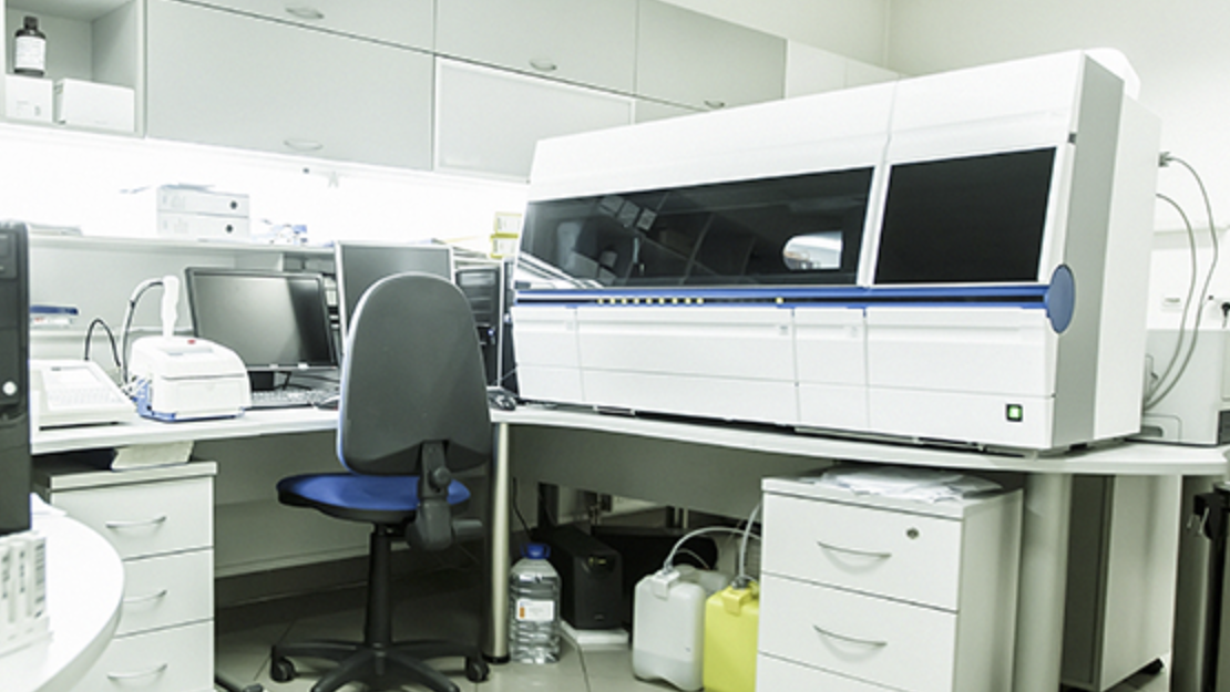
Departmental Research Facilities can be grouped into four groups:
Biomedical Engineering
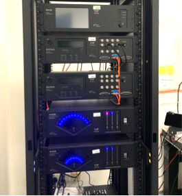
32 Channel Recording System
Description:
The 32 channel recording system is a group of digital signal processors which can be used to acquire, process and analyse multi-site electrophysiological/neural signals. It can also provide real-time assessments and monitoring for animal behavioural experiments, and output customized stimulations to experimental subjects.
Person In-charge:
Prof. Ed Wu
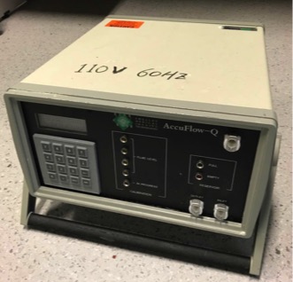
AccuFlow-Q
Description:
The flow pump can generate different types of blood flows, including constant, sinusoidal, carotid arterial and femoral arterial flows at controllable flow rates.
Person In-charge:
Dr. W.N. Lee
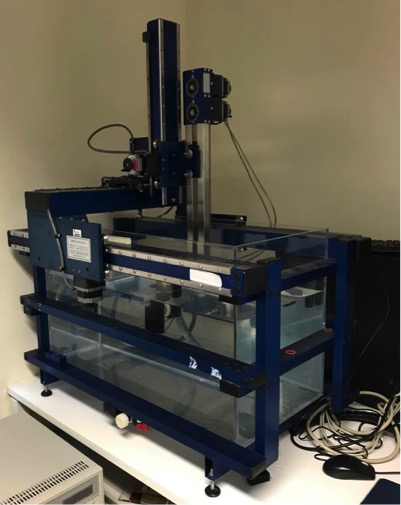
Acoustic Intensity Measurement System (Onda Corporation)
Description:
The 3D positioning system is comprised of a water tank with a positioning controller, a desktop PC with control and processing application software, an oscilloscope, and a function generator for the measurement of acoustic fields. An ultrasound transducer and a hydrophone are placed in the water tank filled with de-gassed water. The 3D positioning system controls the positions of the transducer and the hydrophone.
Person In-charge:
Dr. W.N. Lee
High-field 7-Tesla MRI Scanner
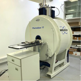
Description:
The 7-Tesla MRI Scanner is a high-field NMR imaging system which offers advanced capabilities to study soft tissue structures and functions in small animals. It consists of an actively shielded 7T horizontal superconducting magnet with a 16 cm bore, gradient control system, RF transmitting and receiving coils, and the computer console.
Person In-charge:
Prof. Ed Wu
Nanowizard4-XP Bio-AFM system
Description:
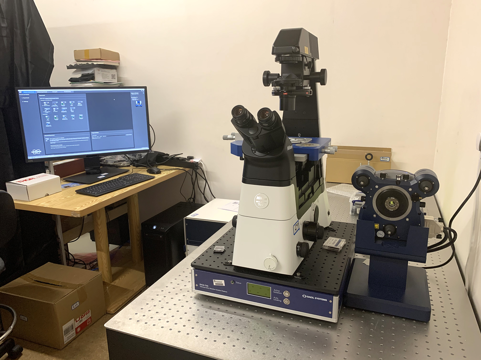
The NanoWizard® 4 XP BioScience atomic force microscope combines atomic resolution and fast scanning with rates of up to 150 lines/sec and a large scan range of 100µm in one system. It is designed to provide highest mechanical and thermal stability on inverted optical microscopes during long term experiments on samples ranging from single molecules to living cells and tissues.
Person In-charge:
Dr. Z. Chu
Verasonics® Vantage 256 System
Description:
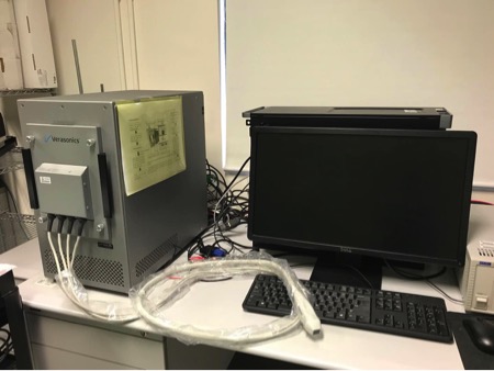
Verasonics system is a fully programmable open architecture ultrasound system. The system is comprised of three main components— a data acquisition module, a host controller and Matlab®-based software package. The programmability through Matlab and full access to the channel data permits to develop advanced imaging algorithms and products in ultrasound research. The system is currently used for B-Mode imaging, Doppler imaging, ultrafast ultrasound imaging, shear wave and strain imaging and other ultrasound imaging research.
Person In-charge:
Dr. W.N. Lee
Ultrasonix® RP System
Description:
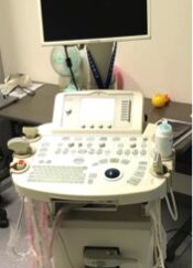
The RP System is a diagnostic ultrasound scanner with both a research and a clinical mode. In the research mode, the system is designed to allow users to pre-define parameters and access raw data (i.e., beamformed radiofrequency (RF) data) that are not provided in other clinical systems. The system offers A-mode, B-mode, M-Mode, power Doppler, pulsed Doppler, and color Doppler.
Person In-charge:
Dr. W.N. Lee
Electrical Energy and Control Systems
32 Channel Recording System
Description:
The 32 channel recording system is a group of digital signal processors which can be used to acquire, process and analyse multi-site electrophysiological/neural signals. It can also provide real-time assessments and monitoring for animal behavioural experiments, and output customized stimulations to experimental subjects.
Person In-charge:
Prof. Ed Wu
AccuFlow-Q
Description:
The flow pump can generate different types of blood flows, including constant, sinusoidal, carotid arterial and femoral arterial flows at controllable flow rates.
Person In-charge:
Dr. W.N. Lee
Acoustic Intensity Measurement System (Onda Corporation)
Description:
The 3D positioning system is comprised of a water tank with a positioning controller, a desktop PC with control and processing application software, an oscilloscope, and a function generator for the measurement of acoustic fields. An ultrasound transducer and a hydrophone are placed in the water tank filled with de-gassed water. The 3D positioning system controls the positions of the transducer and the hydrophone.
Person In-charge:
Dr. W.N. Lee
High-field 7-Tesla MRI Scanner
Description:
The 7-Tesla MRI Scanner is a high-field NMR imaging system which offers advanced capabilities to study soft tissue structures and functions in small animals. It consists of an actively shielded 7T horizontal superconducting magnet with a 16 cm bore, gradient control system, RF transmitting and receiving coils, and the computer console.
Person In-charge:
Prof. Ed Wu
Nanowizard4-XP Bio-AFM system
Description:
The NanoWizard® 4 XP BioScience atomic force microscope combines atomic resolution and fast scanning with rates of up to 150 lines/sec and a large scan range of 100µm in one system. It is designed to provide highest mechanical and thermal stability on inverted optical microscopes during long term experiments on samples ranging from single molecules to living cells and tissues.
Person In-charge:
Dr. Z. Chu
Verasonics® Vantage 256 System
Description:
Verasonics system is a fully programmable open architecture ultrasound system. The system is comprised of three main components— a data acquisition module, a host controller and Matlab®-based software package. The programmability through Matlab and full access to the channel data permits to develop advanced imaging algorithms and products in ultrasound research. The system is currently used for B-Mode imaging, Doppler imaging, ultrafast ultrasound imaging, shear wave and strain imaging and other ultrasound imaging research.
Person In-charge:
Dr. W.N. Lee
Ultrasonix® RP System
Description:
The RP System is a diagnostic ultrasound scanner with both a research and a clinical mode. In the research mode, the system is designed to allow users to pre-define parameters and access raw data (i.e., beamformed radiofrequency (RF) data) that are not provided in other clinical systems. The system offers A-mode, B-mode, M-Mode, power Doppler, pulsed Doppler, and color Doppler.
Person In-charge:
Dr. W.N. Lee